Orthopedics
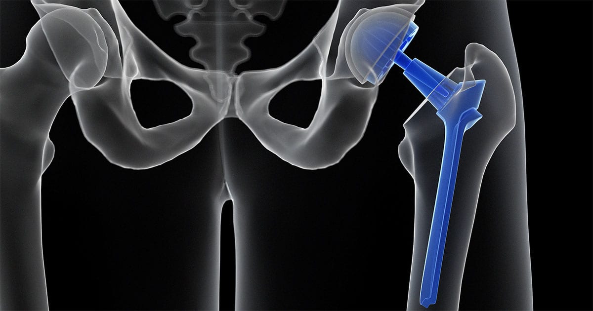
Total hip arthroplasty
day: Monday,31, July,2023
Place: Nariman orthopedic hospital
Time: 9:00 am
Anatomy
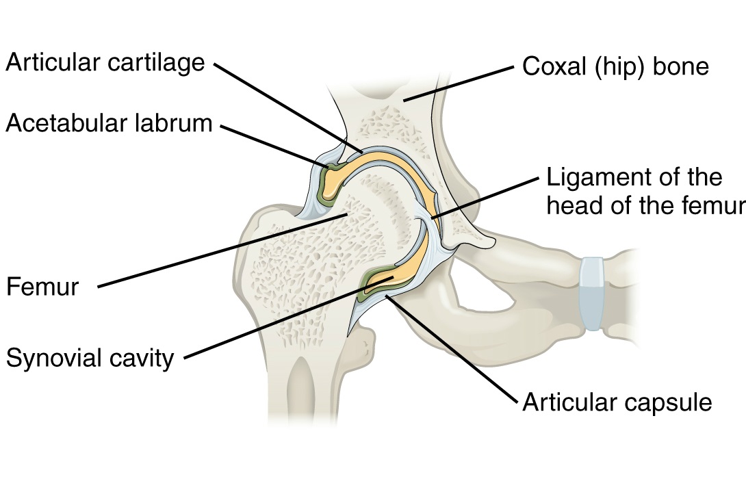
The hip is one of the body's largest joints. It is a ball-and-socket joint. The socket is formed by the acetabulum, which is part of the pelvis bone. The ball is the femoral head, which is the upper end of the femur (thighbone).
The bone surfaces of the ball and socket are covered with articular cartilage, a smooth tissue that cushions the ends of the bones and enables them to move easily.
A thin tissue called the synovial membrane surrounds the hip joint. In a healthy hip, this membrane makes a small amount of fluid that lubricates the cartilage and eliminates almost all friction during hip movement.
Bands of tissue called ligaments (the hip capsule) connect the ball to the socket and provide stability to the joint.
Common Causes of Hip Pain
-Osteoarthritis
-Rheumatoid arthritis
-Posttraumatic arthritis
-Osteonecrosis
Description
During hip replacement, a surgeon removes the damaged sections of the hip joint and replaces them with parts usually constructed of metal, ceramic, and very hard plastic. This artificial joint (prosthesis) helps reduce pain and improve function.


Comments
Post a Comment